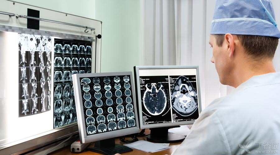
Imaging examination is a common examination method for doctors, including X-ray, ultrasound, CT, nuclear magnetic resonance, etc.
Many patients will have doubts, so many examination methods, why do I need to do this? Why is it that the doctor asked the patient to have an MRI while asking me to have a CT? Is the more expensive the examination, the clearer it will be?
It is precisely because many people have such and such doubts that I am here to talk about it, hoping it will be helpful to you.
How to choose imaging examination?
First, let’s have a brief version, which is not reasonable, but practical.
1. Bones
X-ray films are used for preliminary screening. If the diagnosis is not clear, CT; can be further used. MRI can be used to observe joint soft tissue and bone tumor lesions.
2. Cervical spine, lumbar spine, etc.
For spinal lesions, MRI is the best, followed by CT.
3. Brain and spinal cord
For acute stroke and trauma, CT should be performed first to eliminate hemorrhage. MRI can be used to analyze the severity of stroke in detail. Most of the other brain or spinal cord lesions are best by MRI.
Step 4: Chest
Roughly understand the selection of X-ray films, carefully analyze and select CT, and generally do not select MRI when looking at lungs.
5. Abdomen, pelvic cavity
The parenchymal organs can be treated by ultrasound, and CT and MRI also have their own advantages.
6. Esophagus, stomach, intestinal tract, etc.
Barium meal, i.e. Barium contrast of digestive tract, can be used.
7. Heart
Look at the heart itself with ultrasound. Patients who are not suitable for coronary angiography can choose CT to exclude the possibility of coronary heart disease, and nuclear magnetic resonance can be used as supplementary examination.
The above contents should be used as a reference for choosing the examination method when seeing a doctor. Next, I will talk about it in detail.
What are all these tests?
1. X-ray film
Commonly known as [film]. It is widely used in the examination of spine, limbs, chest and abdomen.
The advantages are fast inspection and low price.
However, because it is a plane overlapping image of human tissue, diagnosis has limitations.
2. Barium Meal
Barium contrast of digestive tract, i.e. X-ray film of digestive tract. By introducing barium, images with obvious differences from human tissues can be produced, enabling doctors to more clearly identify common diseases under the dynamics of gastrointestinal tract.
Is contrast agent harmful to human body?
Contrast agents will be used to help develop the contrast. Generally speaking, people with allergy and high-risk related diseases need special attention. For people without contraindications, it is safe to use them.
3. CT
By using X-ray to scan a certain part of the human body, the cross-sectional image of the examined part of the human body can be obtained, or the stereo image can be generated after processing. Compared with X-ray films, CT has stronger resolution ability and can display soft tissue lesions that cannot be found by conventional X-ray examination.
4. Ultrasound
It can provide anatomical and tissue hemodynamic information.
Ultrasound examination is radiation-free, very safe and even suitable for prenatal examination of pregnant women. However, ultrasound penetration is weak and has limitations in diagnosing some pathological changes.
5. MRI
Magnetic resonance imaging.
The advantages are no radiation, high tissue resolution and multi-type imaging.
Disadvantages include long examination time, loud noise and small space. MRI examination is not recommended for patients who cannot cooperate (such as children, delirious, claustrophobic patients, etc.). In addition, the examination cost of MRI is also relatively high.
It should be noted that MRI-is strictly prohibited for patients with metal implants (such as cardiac pacemakers, cardiac mechanical valves, aneurysm clips, vascular stents, artificial joints, metal internal fixation, cochlear implants, etc.) in their bodies-unless MRI examination is specifically indicated on the implant instructions.
6. PET/CT
In addition to showing tissue structure like conventional CT examination, it can also evaluate the metabolic function of cells.
This examination is of great significance in evaluating myocardial survival and localization of cerebral epilepsy foci. In recent years, it has gradually been widely used in tumor screening and localization.
However, the examination cost is usually relatively high and there is radiation, so reasonable selection should be made according to the doctor’s advice.

What tests will be done for common diseases?
1. Fracture
Suspected trauma injured bone, X-ray plain film is fast and economical, and is the first choice.
For further and more detailed observation, CT can be selected.
MRI has advantages in bone marrow lesions, soft tissue lesions adjacent to bones and hidden fractures.
Ultrasound is not clear enough to distinguish the cortical medulla of bone and is generally not used.
2. Disc disease
For example, cervical spondylosis, lumbar disc herniation, etc., CT is the fastest and most convenient examination method.
If it is necessary to better observe spinal cord nerves, soft tissues, bone lesions, etc., MRI is the best choice.
3. Examination of joints, muscles, adipose tissue, etc.
MRI is the first choice. In addition, MRI is also very accurate in the diagnosis of brain, spinal cord, abdominal and pelvic organs, cardiac macroangiopathy and myocardial infarction.
Because MRI has excellent soft tissue resolution, it can obtain more comprehensive images and facilitate diagnosis.
4. Acute Stroke
CT examination is used to exclude the presence or absence of hemorrhage, which is convenient for early diagnosis. After the disease is stable, MRI can be used for further detailed observation of lesions.
It should be noted that only CT is performed in the early stage and no abnormality is found, which cannot directly rule out the possibility of cerebral infarction.
5. Chest lesions
Roughly examine the heart, aorta, lung, pleura, ribs, etc. and use X-ray chest films. It can be seen that the heart shadow is enlarged, the lung texture is increased, the lung calcification points, aortic nodal calcification, etc.
Compared with X-ray, chest CT examination shows clearer structure, and its detection sensitivity and accuracy for chest lesions are better than those of conventional X-ray chest films, especially for the detection of early lung cancer.
High-resolution CT further increases the resolution of lung observation, which is of great significance for some diseases (such as pulmonary interstitial diseases).
However, the radiation dose of CT examination is higher than that of X-ray examination.
Which tests have radiation?
There is no ionizing radiation in B-ultrasound and MRI. Imaging methods based on X-ray technology, such as X-ray radiography, various contrast examinations, CT, PET/CT, etc., have ionizing radiation.
In medical examination, the use of X-rays is strictly controlled within a safe range, and reasonable examination will not cause obvious damage to human body. However, pregnant women generally do not recommend radiological examination.
6. Examination of abdominal and pelvic organs
B-ultrasound is often used for routine examination of these organs.
Under the examination of experienced ultrasound doctors, ultrasound has a high diagnostic accuracy for diseases in these parts.
However, ultrasound is greatly interfered by gas, so ultrasound diagnosis is not suitable for areas with more gas, such as intestinal tract.
When ultrasound cannot meet the diagnostic requirements, CT or MRI should be performed.
7. Cardiac examination
Cardiac color Doppler ultrasound, as a routine examination of cardiac structure and function, can provide sufficient information and is commonly used.
The gold standard for the diagnosis of coronary heart disease is coronary angiography. However, CT can be used instead for some patients who are not suitable for coronary angiography because of hospitalization and injection of contrast agent into arteries.
Cardiac MRI is the [gold standard] for evaluating the structure and function of the heart, but it is not used as a routine examination method due to high price and long examination time.
Summary
The imaging principles of various imaging examinations are different, and each has its own advantages and disadvantages. There is no imaging technology that can be applied to the diagnosis and treatment of all human diseases.
The imaging examination methods selected are different for different parts and different observation emphases. Doctors will choose one or more suitable examination methods according to specific conditions to achieve the purpose of accurate diagnosis.
This article has passed the peer review of Dr. Clove’s peer review expert committee.
Copyright of Clove Garden. No reprinting is allowed without permission.
