
[Cardiac stent] is the most representative and frequently talked about instrument in contemporary cardiology. Whether praising the advanced modern medicine or criticizing the high cost of medical consumables, we always have to mention this small [stent]. Its appearance has changed the history of cardiology.
However, how many people have seen this familiar instrument and how many people know how it is put into the blood vessels of the heart?
[Stenting] is completed through [interventional surgery], we might as well start with [cardiac interventional surgery].
The Art of Threading Needle and Lead Thread, Let Scalpel Sing Supporting Role
If cardiac surgery is the heroic act of cardiac surgeons, then cardiac interventional surgery is the elegance of cardiac physicians.
Generally speaking, cardiac intervention does not open the belly like traditional surgery, but passes through small catheters and blood vessels to reach the lesion site, thus performing operations to complete diagnosis and treatment.
The principle is not complicated-the main arteries of the human body originate from the heart and gradually divide into various arteries to supply various limbs and organs. After dispersing into small arteries and capillaries, they merge into veins. The veins of the whole body merge again and return to the heart through the main veins. In other words, the blood vessels of every part of the whole body are directly or indirectly connected to the heart.
Therefore, the blood vessels of the human body are the natural channels leading to the heart, and interventional surgery is completed by entering the heart along the blood vessels.
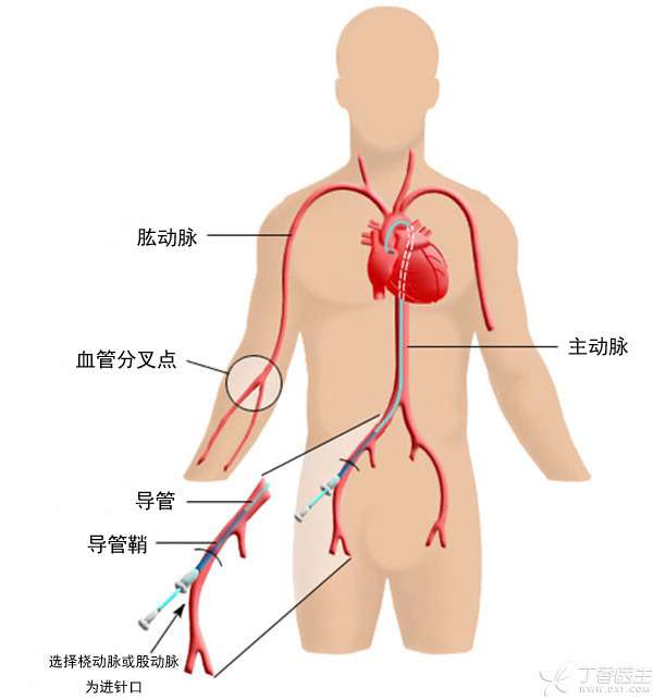
Fig. 1 Blood vessels pass through the heart.
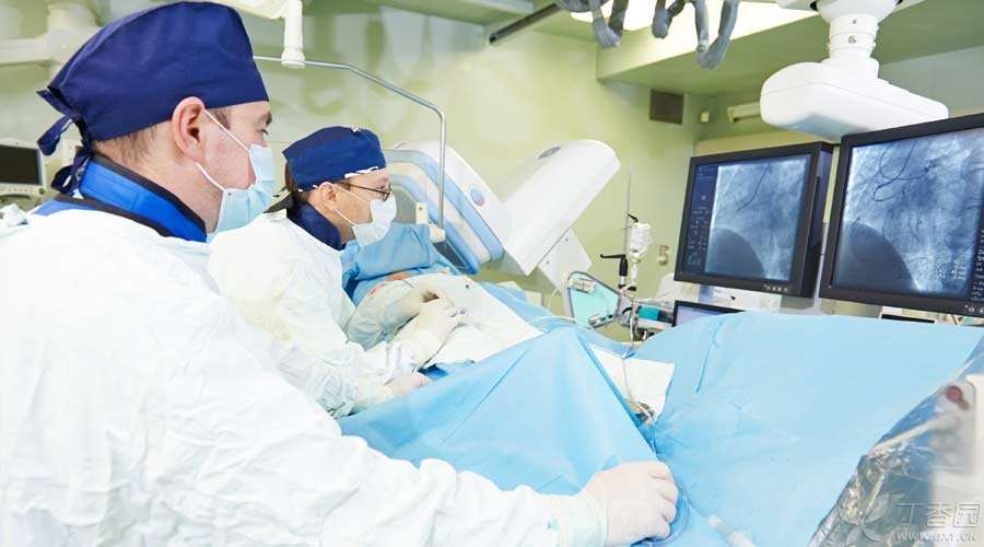
In such operations, the scalpel is no longer the main role, and the surgical operating room is no longer the operating ground. Doctors mainly rely on guidewire and catheter for operation. The operation is often carried out in the [catheter room] specially equipped with fluoroscopy equipment and radiation shielding, and these operations are all completed by cardiologists.
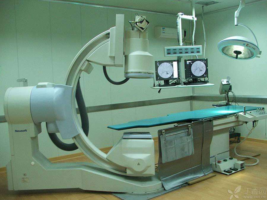
Fig. 2 catheter chamber equipped with fluoroscopy equipment
Minimal trauma, meticulous work, saving tens of millions of lives,
Coronary heart disease is caused by stenosis and occlusion of coronary arteries that supply blood to the heart. Stenting has become one of the mainstream techniques to treat coronary heart disease. Millions of patients worldwide undergo the operation every year. In China, as many as 400,000 cases are carried out every year. What’s more, this kind of operation requires only minimal trauma.
How is this done?
1. Establish [channel]
First of all, we need to establish a channel into the blood vessels. Usually, like injections, special needles are used to directly puncture the blood vessels. The most common choice is the radial artery on the wrist or the femoral artery on the medial thigh.
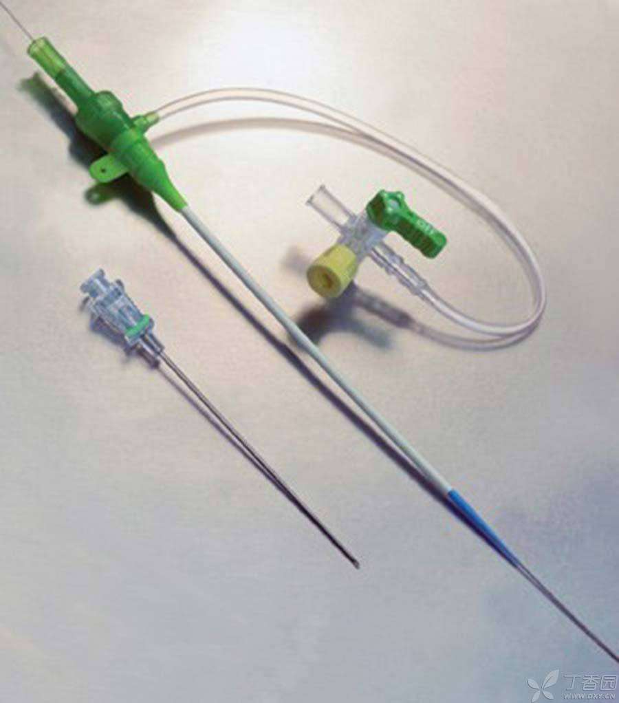
Fig. 3 Needle and sheath used to establish passage in puncture blood vessel
Step 2 Insert the guide wire
Then, the wire is placed along the needle, and the hour hand can withdraw, leaving the wire in the blood vessel. With this wire as the skeleton, the doctor puts the sheath tube and supports the blood vessel out of a channel, so that the guide wire and catheter can go all the way down the channel to the heart until reaching the coronary artery.
The guidewire here is not a normal wire, Instead, it is a very thin and precise material, which is soft as a whole, and the tip can be flexibly bent and turned by the doctor, crossing complicated paths from large blood vessels thicker than the thumb to coronary arteries thinner than noodles. The catheter can meander through the blood vessels along the path of the guidewire.
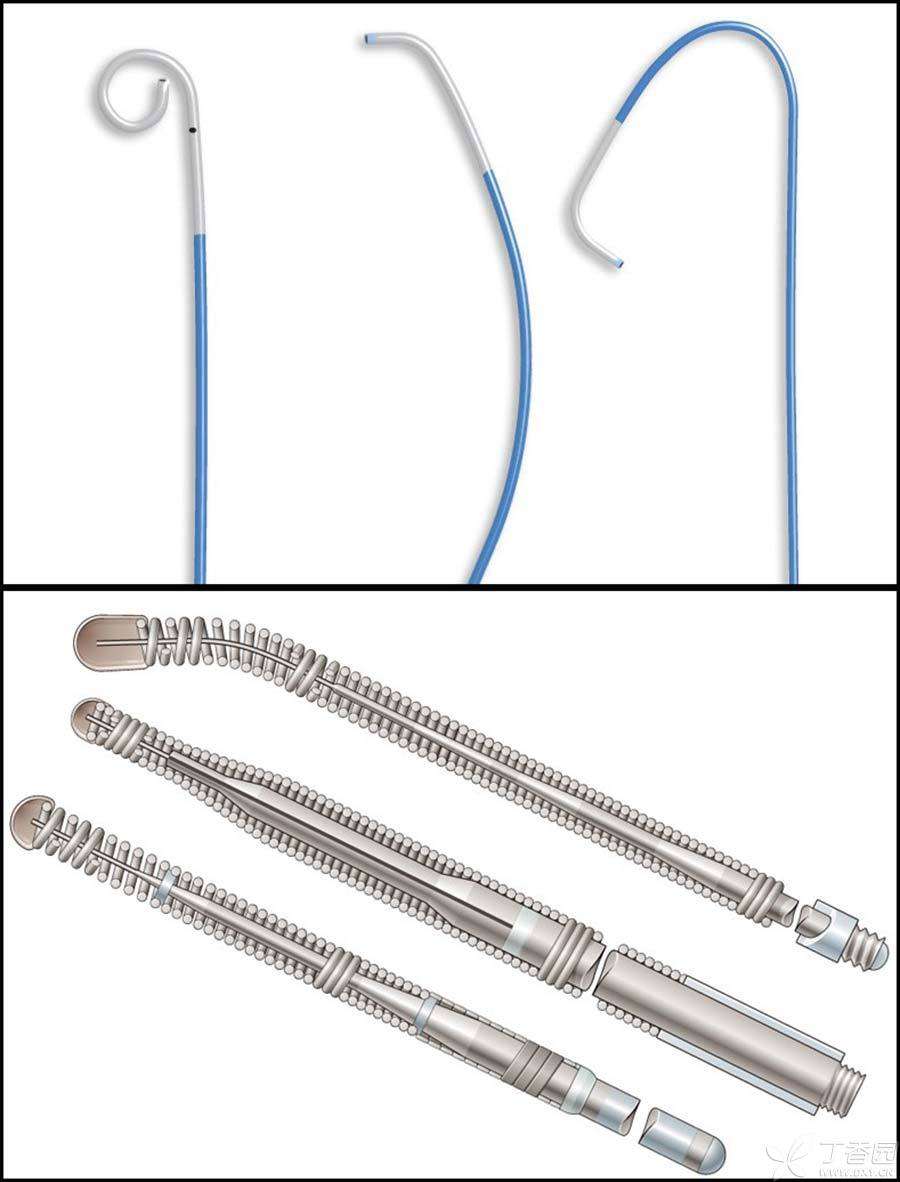
Fig. 4 The catheter and guidewire can freely meander in the blood vessel because the precise guidewire tip can be flexibly deformed and turned under the operation of the doctor.
3. Angiography
In the process of arriving at the destination all the way, doctors need to observe the situation through X-ray fluoroscopy, and also need to push [contrast agent] through catheter to clearly display blood vessels and find the lesion site under X-ray.
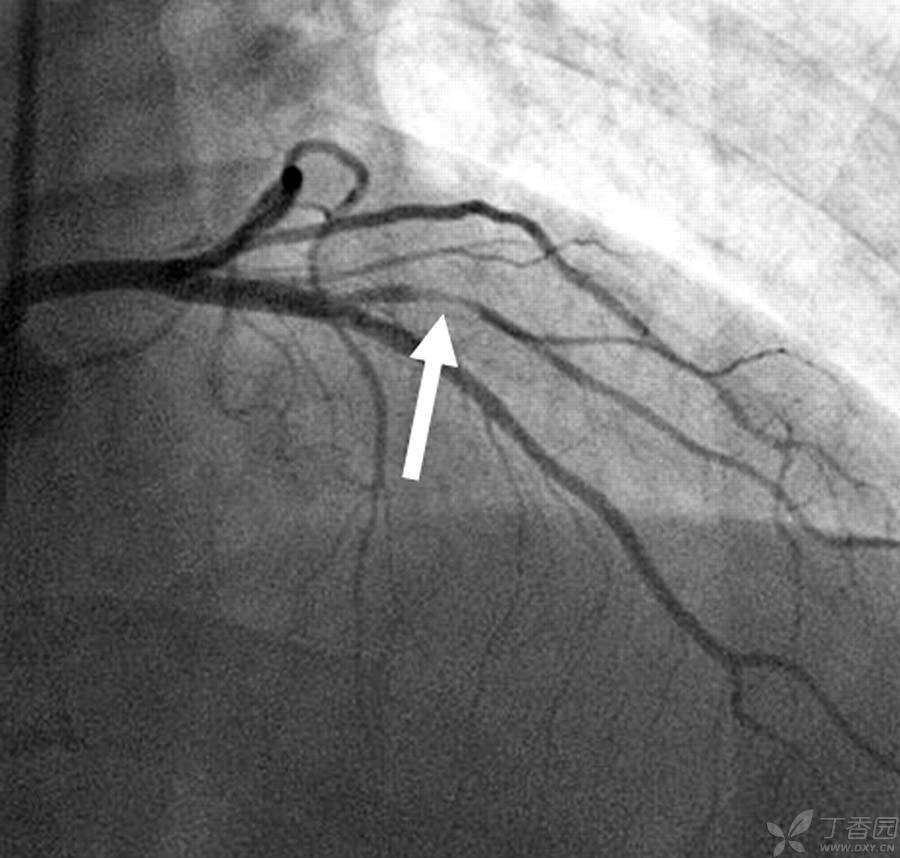
Fig. 5 Coronary artery under X-ray fluoroscopy, arrow shows lesion stenosis blood vessel
4. Insert balloon and stent
Along the catheter, the doctor sent a balloon wrapped with the metal stent into the blood vessel at the lesion site, inflated the balloon, and stretched the metal stent open at a pressure of about 10 times atmospheric pressure.
This kind of support, once placed properly, will maintain its shape for life and permanently support narrow parts.
In order to avoid long thrombosis on bare metal and cause stenosis and obstruction again, the new generation of stents have a layer of drug coating or even biological coating on the surface, greatly reducing the incidence of restenosis.
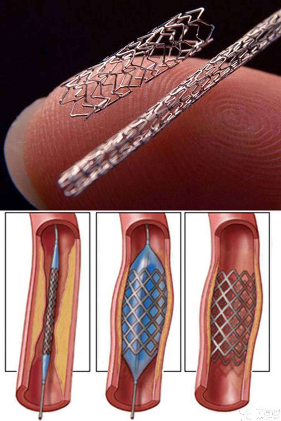
Fig. 6 Opening process of cardiac stent
The above is the main process of cardiac stent placement. Does it not seem complicated or even too simple compared with traditional surgery?
In fact, the above process is often a race against time. Patients have already suffered from myocardial infarction and are in critical condition. They are sent to emergency department. Whether the obstructive blood vessels can be opened as soon as possible is the key to save life and health. Moreover, the actual situation is ever-changing, and the difficulty and complexity of pathological changes are different. Once problems occur in many links, life can be endangered. It is not rare to rescue cardiac arrest in catheter room.
Time-limited homework requires caution and meticulous work. It also requires rich experience and bold decision-making. In fact, the bracket technique of “threading the needle and leading the wire” is a skillful application of a weapon and a set of martial arts, not a bird embroidered in a purdah.
Wearing lead armor to resist radiation, he sacrificed for medicine.
After inserting the catheter into the blood vessel, if there is no pair of fluoroscopic eyes, then the interventional operation is doomed to be blind. Fortunately, X-ray technology has given us such a pair of fluoroscopic eyes, making the extremely small blood vessels clearly visible, making the interventional operation possible. However, the cost is huge.
Every time I use X-ray perspective to observe, it is a radiation release. This is a real X-ray that can cause damage, rather than the innocuous civil electromagnetic waves such as mobile phones and routers. After several operations, the scattering received by standing beside the patient is also considerable.
As a patient, there is no need to worry. A coronary artery operation usually takes only half an hour to an hour. The radiation dose is often within the tolerance range and the health risk is very small. However, as a doctor, as many as hundreds of such operations a year will accumulate considerable radiation dose after decades of work.
In order to reduce radiation damage, doctors need to wear a complete set of protective clothing-lead aprons, vests, belts, scarves, hats, masks, etc., which add up to 30 to 40 kg. Going on the operating table is like wearing an ancient armor.
However, such protection still cannot guarantee the health of the surgeon. Some bodies are always exposed and radiation scattering is everywhere.
According to incomplete statistics, interventional doctors have a higher risk of malignant tumors, cataracts and other diseases than other medical personnel, and joint and spinal problems are also quite high due to long-term heavy lead clothing. Although protection technology is also continuously improving, interventional doctors continue to sacrifice for medicine until the day when the technology is perfect.
The above is the process of putting a small stent into the heart.
In fact, interventional technology can not only save people with coronary heart disease, but also implant permanent pacemakers through interventional technology to treat arrhythmia and heart failure, congenital heart disease and valvular heart disease, etc. It is already a routine project. The rapid development of medical technology will open your eyes every few years.