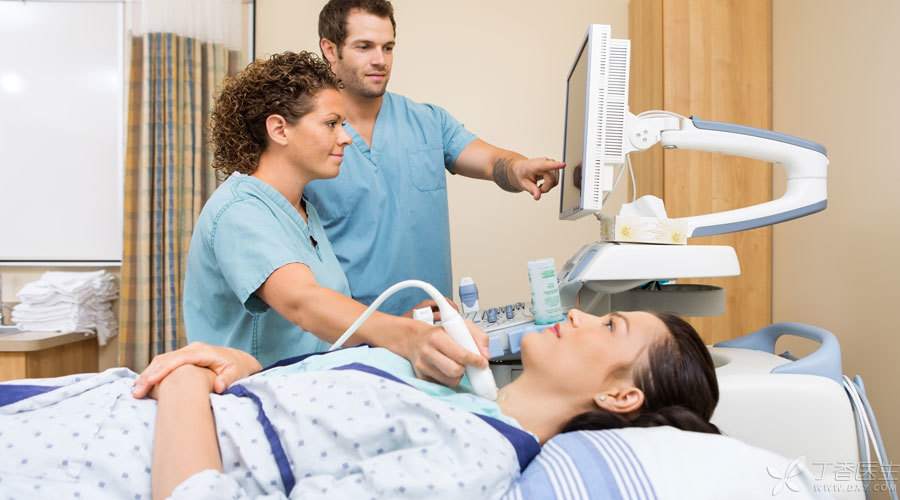
When examining the thyroid gland, besides blood tests, thyroid ultrasound is an indispensable item. Ultrasound can be divided into black-and-white ultrasound and color ultrasound, just like TV has black-and-white and color difference? No. On the monitor, Color ultrasound also sees black and white images. The reason why it is named [color] is that it can display the blood flow of the inspected part, and the flow direction is different, and the blood is [given] different colors, so the whole color ultrasound is called [color Doppler blood flow imaging], abbreviated as [color Doppler ultrasound]. In addition, it is clearer and has better image quality than black and white ultrasound.
Usually used for thyroid examination is mostly color Doppler ultrasound. Why? The thyroid gland is just below the muscle, The position is not deep, The blood flow is very rich, It is most suitable to use color Doppler ultrasound for examination. The doctor will record the examination contents into words in sequence. Intercept the characteristic image again, finally give the diagnosis opinion, a thyroid color Doppler ultrasound report is formed. Look at the picture is a doctor’s thing, the text content may be what you want to know most. It is better to follow the color Doppler ultrasound to a real combat, and look at the process of a thyroid color Doppler ultrasound report.
Step 1: Determine the location
Description of the report: Thyroid anatomy is normal.
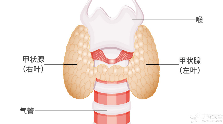
Interpretation: Under normal circumstances, the thyroid gland always installs its home between the 5th cervical vertebra and the 1st thoracic vertebra, roughly 2 cm below Adam’s apple. The anatomical position of the thyroid gland is normal, indicating that the thyroid gland is in the normal position.
Not all people’s thyroid gland will obediently grow in this position, a few people because of congenital factors, thyroid gland may settle in another country, such as the root of the tongue, or behind the sternum, this is called ectopic thyroid gland. Ectopic thyroid gland to grow from the root of the tongue to the normal position of thyroid gland is more common.
If the thyroid gland is not found in the normal position, further CT and nuclide scanning may sometimes be required.
Step 2: Pay Attention to [Body]
Description of the report: Thyroid morphology is abnormal, the left lobe is 68 × 29 × 23 mm in size, the right lobe is 63 × 28 × 22 mm in size, and the isthmus is 8 mm in thickness. The capsule is smooth and the boundary is clear.
Interpretation: The normal [body] of thyroid gland is [H] type, the [,] on both sides are the left and right lobes of thyroid gland, and the [-] in the middle is the isthmus of thyroid gland. The size of the left and right lobes is recorded by height × width × thickness, and the corresponding normal value is (45 ~ 60 mm) × (15 ~ 25 mm) × (15 ~ 20 mm). In most cases, the size of the two lobes is symmetrical, and the asymmetrical reasons may be surgery, congenital dysplasia, etc. The thickness of isthmus was recorded, and the normal value was 2 ~ 6 mm.
According to this standard, the height, height, weight and thinness of thyroid gland are expressed as normal, swollen and reduced. The thyroid gland in the report is swollen.
Goiter is more common in hyperthyroidism, Hashimoto thyroiditis and nodular goiter, some acute or subacute thyroiditis, etc. Thyroid reduction is found in the late stage of Hashimoto’s thyroiditis after iodine 131 treatment. In addition, congenital developmental abnormalities and thyroid reduction will also occur after partial thyroidectomy.
Thyroid gland is also wearing a coat, it is the envelope of thyroid gland. Normal thyroid gland or pathological changes do not affect the envelope of thyroid diseases, envelope and the outside boundary is clear. Boundary blurred, indicating inflammatory or tumor diseases, some pathological changes even break through the thyroid envelope, implicated in thyroid tissue around the trachea, nerve tissue, etc.
In case of the above abnormalities, further examination of thyroid function, thyroid autoantibodies, etc. is required.
Step 3: Evaluate [Yan Value]
The report describes: The thyroid parenchyma echo is uneven. Multiple hypoechoic areas can be seen in the left lobe with regular edges, uniform internal echoes and clear boundaries, with the largest being about 0.6 × 0.4 cm, while anechoic areas can be seen in the right lobe with clear boundaries, with a size of about 1.0 × 0.3 cm.
Interpretation: Under the coat of the thyroid gland, the body-substantial part of the thyroid gland is wrapped. Color Doppler ultrasound emits ultrasonic waves, which are reflected back to form images on the display.
1. Is the echo uniform
If the reflected echo is uniform, the thyroid substance seen is also flat and smooth, and the [color value] is excellent.
If the reflected echo will be uneven, it indicates that the substance is uneven, like the surface of the moon, indicating diffuse thyroid diseases, such as hyperthyroidism, Hashimoto’s thyroiditis, etc.
2. Echo intensity
If the echo intensity is reduced, it indicates that the essence has been damaged, which is found in Hashimoto’s thyroiditis, subacute thyroiditis and other diseases.
Increased echo intensity is rare and can be seen in thyroid nodules.
3. Thyroid nodules
If the thyroid gland grows nodules, It’s like pimples on your face, [Yan Value] has been discounted again. Color Doppler ultrasound should not only be from the outside, The number of nodules (single or multiple), size, shape (oval, round-like, irregular), boundary (boundary between nodules and surrounding normal thyroid tissue), Clear or fuzzy), whether the edge is smooth and complete, etc., also want to distinguish solid, cystic or cystic solid nodule from the internal structure of the nodule. Solid nodule refers to the nodule is completely filled with solid matter, cystic nodule can be liquid or hollow. Cystic solid nodule refers to the nodule is cystic and mixed.
How to distinguish clearly?
Ultrasound scans the nodules, There is also reflected echo. According to the echo intensity, it can be divided into anechoic, hypoechoic, isoechoic and hyperechoic. Ultrasonic waves have echoes when encountering solids. And the solid density is different, echo has low, equal and high difference. However, ultrasonic wave meets liquid or hollow situation, is unable to reflect echo. Therefore, the report suggests that nodules have no echo, generally cystic nodules, and low, equal or high echo is solid nodule.
Like the thyroid parenchyma, there are reflective echoes inside the nodule, which can be divided into uniform or uneven echoes. When calcium salts in the body deposit in the nodule, calcification can occur, which is shown on color Doppler ultrasound and described as strong echo. According to the shape, there are tiny or sandy calcification, coarse calcification and annular calcification.
Color Doppler ultrasound is one of the methods to evaluate benign and malignant thyroid nodules.
If the following two conditions are seen in the ultrasound report, the nodule may be benign in more than 90%: ① pure cystic nodule; (2) More than half of the volume of nodules is occupied by multiple small vesicles, showing spongy changes. However, the more the following words appear in the report, the greater the possibility of malignant nodules is: ① solid hypoechoic nodules; (2) Abundant blood flow in nodules, especially under normal thyroid function; (3) The shape and margin of nodules are irregular; ④ Microcalcification, needle-like diffuse distribution or cluster distribution of calcification in nodules; ⑤ At the same time, it is accompanied by abnormal cervical lymph nodes, such as round lymph nodes, irregular or fuzzy boundaries, internal calcification, etc.
For thyroid gland with [color value] problems, thyroid function, thyroid peroxidase antibody, thyroglobulin antibody, etc. should be checked. If malignant is suspected, thyroid biopsy can be performed.
The blood flow inside the nodule is mentioned above-being able to detect blood flow signals is the advantage of color Doppler ultrasound. This is the next step to be taken.
Step 4: Detect Blood Flow
The report describes: Thyroid blood flow signals are abundant, and some blood flow signals can be seen at the edge of nodules.
Interpretation: Color Doppler ultrasound gives color to flowing blood, “that blood flow to the color Doppler ultrasound probe is red on the display, Blood flowing away from the probe is shown as blue, which is called blood flow signal. Therefore, on the image reported by color Doppler ultrasound, two colors can be seen: red and blue. If the thyroid color Doppler ultrasound sees dense red and blue colors, it looks like a sea of fire, which will be described in the report as “fire sign” and is generally found in hyperthyroidism patients.
In addition, blood flow signals of thyroid nodules need to be detected. Blood flow signals can appear inside or at the edge of thyroid nodules. Malignant nodules rely on blood supply to grow, so blood flow signals are common in malignant nodules.
Thyroid function needs to be checked if there is fire sign or rich blood flow. Suspected malignant, may need puncture or surgery.
Step 5: Score
Description of the report: Bilateral thyroid multiple nodules, TI-RADS grade 2.
Interpretation: When you get the thyroid color Doppler ultrasound report, you will usually see the word [TI-RADS classification]. Is this what?
In fact, [TI-RADS] is a thyroid image reporting and data system. According to this system, doctors can grade and diagnose nodules found by thyroid ultrasound-the higher the grade, the greater the possibility of malignant nodules (i.e. Thyroid cancer). Generally, TI-RADS classification is divided into 6 grades:
- Grade 0: No nodule (malignant possibility is 0%), normal thyroid gland or only swollen thyroid gland; Grade 1: Most cases are benign nodules (malignant possibility is 0% ~ 7%); Grade 2: Some possible benign nodules (malignant possibility is 7% ~ 23%); Grade 3: The benign and malignant nodules cannot be determined (the possibility of malignant nodules is 24% ~ 50%);
For patients with grade 3 and below, benign nodules may be larger. If patients have no clinical symptoms, they can be observed regularly, and color Doppler ultrasound will be reexamined for comparison within 3 months to 6 months. If the nodule is large, swallowing and dyspnea caused by compression of trachea and esophagus can be treated surgically.
- Grade 4: malignant may be relatively high, with a possibility of 51% ~ 90%; Grade 5: highly suspected to be malignant, with a malignant possibility of 91% ~ 100%.
Grade 4 patients have the possibility of malignancy, and thyroid biopsy is feasible to determine the nature of nodules.
Grade 5 patients have a great risk of malignancy and may be considered for surgical treatment.
Of course, these are all some [book] knowledge, specific examples, how to do it, need to consider more issues, such as the age of the patient, physical condition, the impact of other diseases, etc.