

Many friends will be confused:
Heart discomfort, how does the doctor judge the specific reason?
Why is it all chest tightness, he can do a color Doppler ultrasound, I have to do contrast?
What can these inspections do separately?
In fact, with the development of technology, there are more effective means to diagnose heart diseases.
Usually, doctors use one or more of cardiac color Doppler ultrasound, electrocardiogram and coronary angiography to help determine what problems in the heart.
Cardiac color Doppler ultrasound to examine cardiac structure
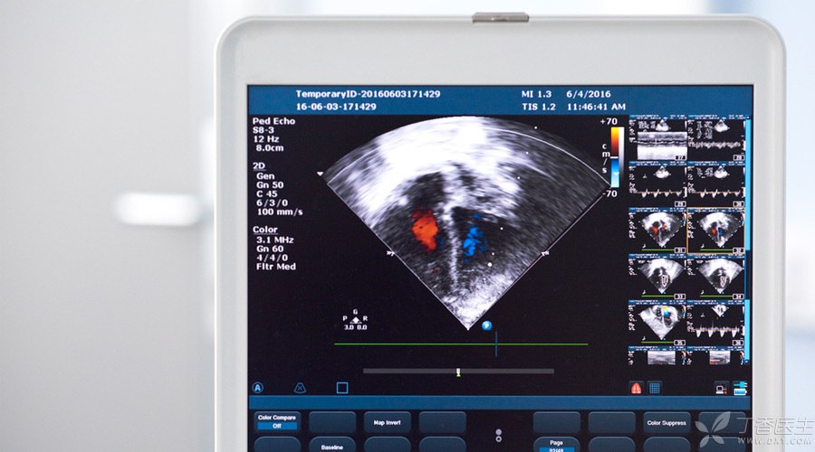
Cardiac color Doppler ultrasound, also known as [heart color Doppler], can dynamically display the heart structure, heart beat and blood flow, and it is very convenient to start examination without any damage.
The heart has four rooms-left and right atria and left and right ventricles. The rooms are separated by [walls] (myocardium). Doors (valves) are the channels through which blood passes through the rooms.
The size of the door and the thickness of the wall have a certain normal range.
Cardiac color Doppler ultrasound can clearly find structural abnormalities of the heart, such as the size of the chamber, the thickness of the septal wall, whether there is insufficiency, stenosis and prolapse of the valve, and the ability of the heart to pump blood.
When some children are born, there are holes in the walls of the room-atrial septal defect and ventricular septal defect, which are often referred to as congenital heart disease.
For patients with hypertension, due to long-term high pressure load from the outside world, the myocardium becomes fatter and the walls become thicker and thicker. This is hypertensive heart disease.
Some people suffer from infection, which leads to rust in the hinges of the door-stenosis or incomplete closure of the valve, which leads to the door not being opened and closed smoothly and affects the normal flow of blood. This is valvular heart disease.
The ultrasonic probe is like the lens of a camera. Moving back and forth in front of the chest can clearly display the various structures of the heart on the screen, and related diseases have no hiding place in front of cardiac color Doppler ultrasound.
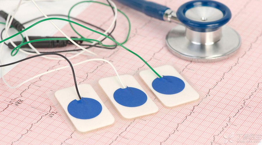
Electrocardiogram, Understanding Heart Rhythm
The heart of a normal person beats 60-100 times per minute. Generally speaking, bradycardia is lower than 60 times and tachycardia is higher than 100 times.
In addition to the number of times, the rhythm of the heart beat is also very important.
The heartbeat of a normal person will be as accurate as a clock. If it is fast and slow, it is often called arrhythmia. Arrhythmia is likely to affect the blood supply capacity of the heart.
For example, atrial fibrillation, the most common clinical arrhythmia, brings the feeling of palpitation, chest tightness, shortness of breath, fatigue and fatigue, and even dizziness and fainting.
ECG can be divided into conventional ECG and dynamic ECG.
The former is to stick the electrode piece and the hand and foot electrical lead system on the skin for a few minutes, and the instrument will use curves to draw the electrical activity of the heart of the examinee. Experienced doctors can know whether the heart beats rhythmically according to these curves.
The advantage of dynamic electrocardiogram is that it can monitor the heartbeat 24 hours a day. After all, arrhythmia is often accidental. Conventional electrocardiogram fails to capture [abnormalities] in time. Dynamic electrocardiogram relies on all-weather [squatting] and can often catch a current situation.
Electrocardiogram is combined with troponin and myocardial enzyme examination, but it is often used to find out the terrorist myocardial infarction!
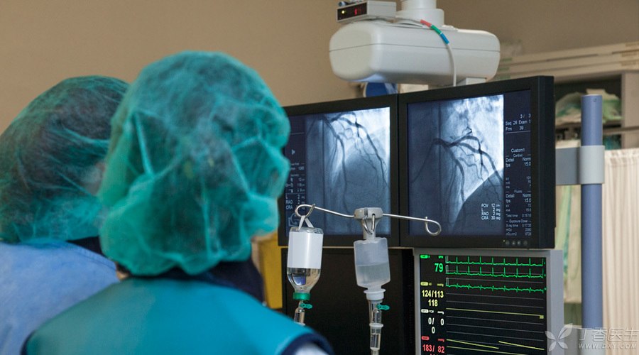
Coronary angiography showing vascular condition
If the heart is a house, then the coronary artery is the pipeline that supplies water and oxygen to the house.
Coronary arteries are distributed outside the heart, providing a steady stream of oxygen and nutrients for the heart.
Diabetes, hypertension, dyslipidemia, smoking, etc., will make more and more impurities in blood vessels and the water flow will become more and more unsmooth.
Generally speaking, if the water pipe is blocked by more than 50%, the body will appear various clinical symptoms with chest pain as the main manifestation under the condition of excitement and tiredness. This is coronary heart disease. At this time, it is necessary to slow down the deposition of impurities in blood vessels through drugs and improvement of lifestyle.
If it is blocked by more than 75% and chest pain and chest tightness are obvious, it may be necessary to perform coronary stent implantation-to stretch the blocked blood vessels with stents or coronary artery bypass grafting-to take a section of blood vessels or blood vessel substitutes from other parts of the body so that blood can bypass the narrow part of the new pipeline and reach the ischemic part.
What examination to know coronary artery condition? It’s coronary angiography.
It is currently the [gold standard] for the diagnosis of coronary heart disease. Through contrast agent, doctors can clearly see the thickness of coronary arteries under X-ray to determine the subsequent treatment.

After reading the article, have you got a better understanding of cardiac examination?
To sum up, the heart structure mainly depends on color Doppler ultrasound, the heart beating rhythm mainly depends on electrocardiogram, and the examination of blood vessels can be realized by coronary angiography.
The doctor examined that with these three treasures, heart diseases have nowhere to run.