
Ultrasound is energy in the form of sound waves. The most common ultrasonic examination is called two-dimensional ultrasound. This type of ultrasound, It is a sensor (commonly known as an ultrasonic probe) that transmits sound waves to the human body. Once these sound waves encounter human tissues, Reflections will occur, just like echoes. After that, sensors will receive these reflected waves. Different human tissues will receive different reflected waves. Through the analysis of these reflected waves by relevant instruments, images of internal organs can be seen. When used on pregnant women, fetuses can be seen.
In gynecology, ultrasound examination is a very important examination and is of great significance in the diagnosis of some diseases, such as pelvic foreign bodies, uterine diseases, pelvic pain or infertility.
Ultrasound examination is more important in pregnancy. According to the requirements of routine pregnancy examination in our country, pregnant women will undergo at least 3-4 ultrasound examinations during the whole pregnancy.
In the early stage of pregnancy, doctors can use ultrasound to eliminate ectopic pregnancy and determine the number of fetuses. In the second and third trimester of pregnancy, the body structure of the fetus can be checked to judge whether the growth of the fetus is normal, determine the orientation of the fetus, observe the respiration and heart rate of the fetus, and also check whether the accessory structures of the fetus such as placenta, umbilical cord, amniotic fluid, etc. are abnormal.
Ultrasound is also used to screen for some congenital diseases, such as Down’s syndrome. In addition, chorionic villus sampling and amniocentesis can also be completed with the help of ultrasound.
Types of Ultrasound Examination
Gynecological ultrasound examination has transabdominal ultrasound and transvaginal ultrasound. According to different needs, doctors decide which type of examination to take.
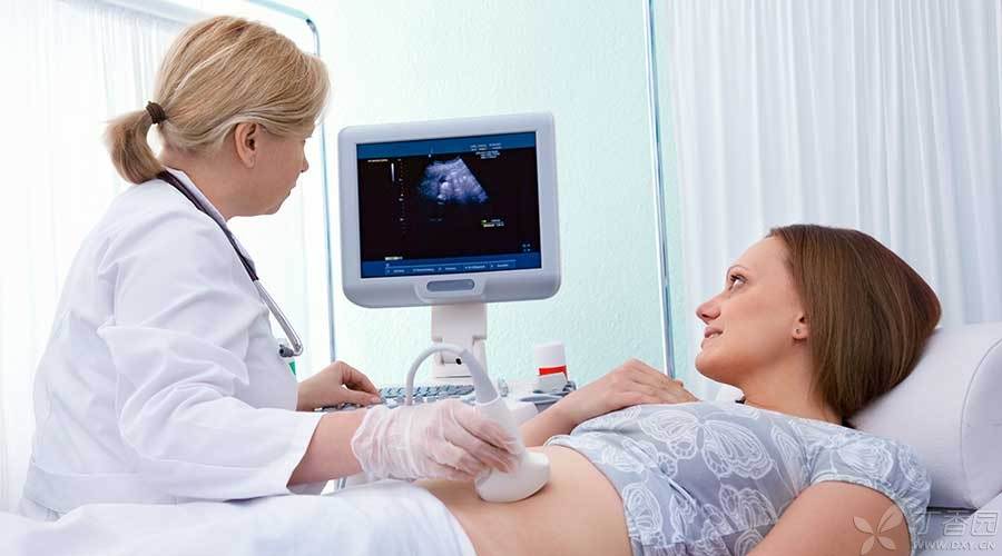
During transabdominal ultrasound examination, Wear loose clothes as much as possible, It is more convenient to expose the abdomen. Ultrasonic examination through the abdomen generally requires holding urine to fill the bladder so that doctors can better observe the ovaries, uterus and other organs of the pelvic cavity. During the examination, doctors will require the entire abdomen to be exposed and apply a gel on the skin that is conducive to sound wave transmission. Doctors will hold the probe and move in the abdomen for examination.
Transvaginal ultrasound does not require holding urine. Its probe is rod-shaped. During the examination, doctors will put a disposable latex sleeve on the surface to prevent the spread of pathogens.
For pregnant women, transvaginal ultrasound is commonly used in early pregnancy, and transabdominal ultrasound is commonly used after 10 weeks of pregnancy.
What is Doppler ultrasound?
This examination is performed during pregnancy by transabdominal ultrasound. Sound waves are used to calculate the blood flow in the umbilical cord or other blood vessels of the fetus, and are also used to listen to the fetal heart.
What is three-dimensional and four-dimensional ultrasound?
Three-dimensional ultrasound is actually a combination of many two-dimensional images taken from many different angles. Four-dimensional images are similar to three-dimensional images, except that they can show the dynamic performance of the fetus. During pregnancy, three-dimensional ultrasound is only used to eliminate fetal abnormalities.
Is the examination to exclude fetal malformation what? Click to view: B ultrasound [large malformation screening]
At present, the [four-dimensional ultrasound] promoted by many commercial organizations does not have greater advantages in determining fetal health, and is mostly aimed at commercial interests.
At present, there is no evidence to prove that ultrasound is harmful to fetal growth, and there is no connection between ultrasound examination and congenital defects, childhood tumors or developmental disorders. Therefore, ultrasound examination is used as the main imaging examination method during pregnancy.
However, some doctors are worried that there may be risks that have not been discovered, so it is not recommended for pregnant women to undergo frequent ultrasound examination without any disease, nor is it recommended for non-medical [four-dimensional] ultrasound.
Responsible Editor: Chuyang